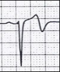
Inverted T Wave On Ecg Causes
The T Wave Physiology Variants And Ecg Features Ecg Echo
T Wave Litfl Medical Blog Ecg Library Basics
T Waves In Ischemia Hyperacute Inverted Negative Wellen S Sign De Winter S Sign Ecg Echo
T Wave Wikipedia

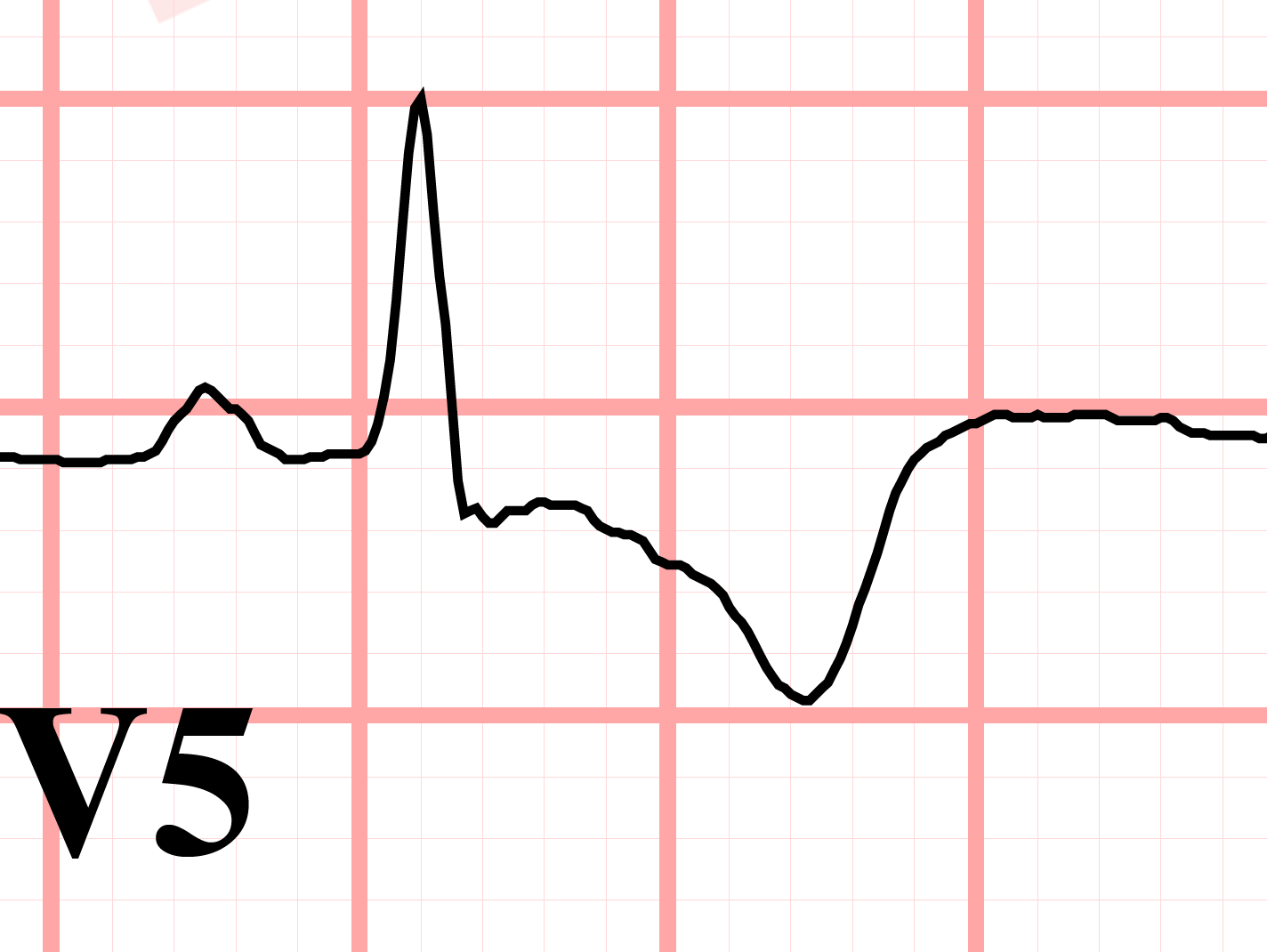
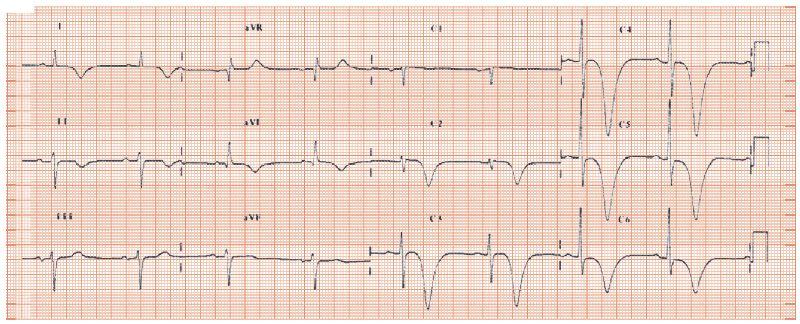
T Wave Inversion In Leads V1 V6 In A 38 Year Old Symptomatic An Download Scientific Diagram

Causes Of T Wave Inversion In Ecg Cardiology And Ccu Facebook
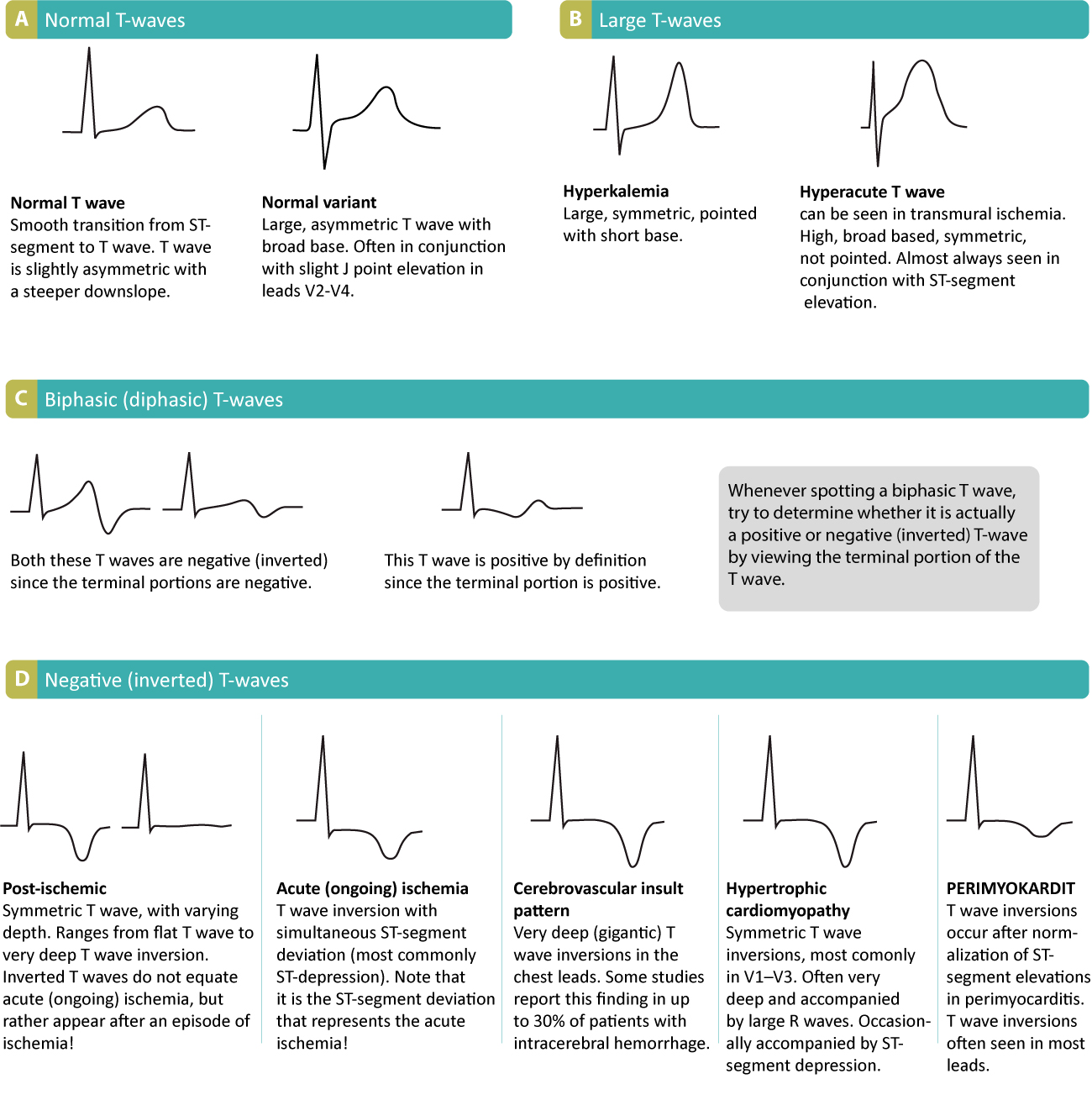
Thus t wave inversions in leads v1 and v2 may be fully normal.
Inverted t wave on ecg causes.
Lesson Title The T Wave
What Is T Wave Inversion Quora
Deep T Wave Inversion Thoracic Key

Mechanism Of Ischemic T Wave Inversion Youtube
Source : pinterest.com
